| Accession number: | 10002 | |
| VFB id: | FBbt_00100484   | |
| Neuron name: | LC12 |
| Synonyms: | |
| Position of cell bodies: | LCBR-LV |
| Number of cells: | 30-35 |
| Neuron class: | visual projection neuron, lobula columnar neuron |
| Innervating regions: | LO3, LO4, PVLP (GL7) [vlpr in Otsuna and Ito (2006), Fischbach and Dittrich (1989)] |
| Presynaptic sites: | PVLP (GL7) [vlpr in Otsuna and Ito (2006), Fischbach and Dittrich (1989)] |
| Postsynaptic sites: | |
| Direction of information: | centripetal |
| Laterality: | ipsilateral |
| Publications: | -
Otsuna and Ito (2006) J. Comp. Neurol. 497:928-958
-
Fischbach and Dittrich (1989) Cell Tissue Res. 258:441-475 |
| |
| Strains / Antibodies: | |
| |
| Morphological description: | LC12 VPNs project to the lateralmost area of the vlpr (Fig. 1). We found 30–35 cell bodies located in the anterior ventral area of the lateral cell body region. The cell body fibers run toward the lobula neck and bifurcate there to contribute to the lobula and to the vlpr. The arborizations in the lobula are thin and columnar.
Synaptic varicosities are observed only in the LO3 and 4 layers (Fig. 1, the left panel). Arborizations from different cells collectively cover all the visual field. At the neck of the lobula, the fibers converge to form a bundle, which projects to the vlpr. Upon entering the central brain, the bundle spreads to form the terminal arborization that has a spherical shape with a diameter of about 15 μm (Fig. 2). The fibers of the LC12 are very short; the length of the fibers between the lobula and the projection target in the vlpr is only about 13 μm, which is the shortest among the 14 lobula-specific VPNs we analyzed. Presynaptic sites are localized only in the vlpr (Fig. 2). Thus, it is likely that LC12 cells are centripetal neurons, which send visual information unidirectionally from the lobula layer LO3/4 to the lateralmost area of the vlpr.
(Otsuna and Ito, 2007) |
| |
| Functional description: | |
| |
| |
| Figure 1: | 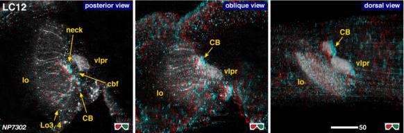 |
| 3D stereograms from three viewing angles. (Neurons other than LC12 are erased from the data.)
CB, position of the cell bodies; neck, medial edge of the lobula where all the lobula-specific VPNs converge; vlpr, ventrolateral protocerebrum; LO3, 4, arborized layers in the lobula. Modified from Otsuna and Ito (2006).
|
| |
Figure 2: | 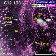 |
| Distribution of the presynaptic sites. Staining with the presynaptic site-targeted n-syb::GFP (green to white) and cytoplasmic DsRed (magenta). White characters with under bar indicate the areas of arborizations with presynaptic sites. Modified from Otsuna and Ito (2006). |
| |
File 1: |  |
| QTVR movie of LC12, from Otsuna and Ito (2006). |