| Accession number: | 10011 | |
| VFB id: | FBbt_00100483   | |
| Neuron name: | LC11 |
| Synonyms: | |
| Position of cell bodies: | LCBR-LDP |
| Number of cells: | 36-40 |
| Neuron class: | visual projection neuron, lobula columnar neuron |
| Innervating regions: | LO2, LO3, LO4, PVLP (GL5) [vlpr in Otsuna and Ito (2006), Fischbach and Dittrich (1989)] |
| Presynaptic sites: | PVLP (GL5) [vlpr in Otsuna and Ito (2006), Fischbach and Dittrich (1989)] |
| Postsynaptic sites: | |
| Direction of information: | centripetal |
| Laterality: | ipsilateral |
| Publications: | -
Otsuna and Ito (2006) J. Comp. Neurol. 497:928-958
-
Fischbach and Dittrich (1989) Cell Tissue Res. 258:441-475 |
| |
| Strains / Antibodies: | NP3045-Gal4 |
| |
| Morphological description: | LC11 neurons have their cell bodies in the posterior dorsal area of the lateral cell body region. We identified 36-40 cells of this category. The cell body fibers run towards the lobula neck and bifurcate there. Projections to the lobula are columnar and form varicosities in two layers. The lateral arborization occurs in the LO2, 3 layers and the lateral area of the LO4 layer, whereas the medial arborization is observed in the medial area of the LO4 layer. The LC11 neurons collectively cover all the visual field.
The fibers form a thin tightly packed bundle at the lobula neck and enter the plpr. The bundle then makes a round turn towards the anterior, without arborization, and enters the vlpr from its posterior border. There, the bundle becomes thicker and make a steep turn laterally to form a stick-like structure (white arrows in Figures 1, 2), which runs anterior-laterally and forms a blunt end in the posterior lateral vlpr.
The strain NP3045 labels only LC11 neurons in the optic lobe. NP7226, on the other hand, labels additional cell cluster of about 15 cells near the LC11 cell bodies. These neurons do not arborize in the optic lobe and project directly to the vlpr (vlpr intrinsic 11: VLPRI11 neurons). They also form a stick-like bundle that lie dorsal ventral to the “stick” component of the LC11 terminals (Figures 2, 3). Unlike LC11, the VLPRI11 neurons form extensive branches that fan out above and below the stick and also in the anterior lateral vlpr (“f” in Figure 3).
The labeling of the n-syb::GFP in the LC11 neurons was observed not in the lobula but only in the “stick” component in the vlpr (Figure 3). The n-syb::GFP signals are densely packed in the whole cross section of the stick. The LC11 neurons are therefore centripetal, sending information from the lobula to the vlpr. On the other hand, the stick component of the VLPR11 neurons is devoid of the n-syb::GFP signal. Presynaptic sites are distributed only in the extensive fan-like braches.
Both LC4 and LC11 have extensive columnar arborizations in the lobula. Their varicosities overlap in LO2 and LO4 layers. Yet, their projection targets in the vlpr are strikingly different. Terminals of LC11 are located relatively dorsally, whereas LC4 terminate more ventrally. LC4 enters the vlpr from its lateral side and terminate in the posteriormost area. LC11, on the other hand, enters the vlpr from the posterior side and arborizes in the lateral and anterior areas. These suggest that apparently similar set of information from the lobula is conveyed to different subregions of the vlpr.
(Otsuna and Ito, 2007) |
| |
| Functional description: | |
| |
| |
| Figure 1: | 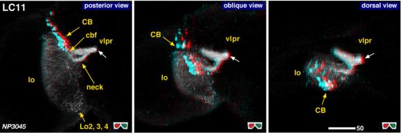 |
| 3D stereograms from three viewing angles. (Neurons other than LC11 are erased from the data. ) White arrow indicates the point where the LC11 bundle makes a round turn.
CB, position of the cell bodies; cbf, cell body fibers; neck, medial edge of the lobula where all the lobula-specific VPNs converge; LO2, 3, 4, arborized layers in the lobula. Modified from Otsuna and Ito (2006).
|
| |
Figure 2: | 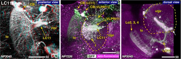 |
| Detailed arborization patterns of LC11. Dashed lines indicate the contour of the vlpr. White arrow indicates the edge of the stick-like arborization. “f” in the middle panel indicates the fan-like projections with scattered varicosities that belong to the vlpr intrinsic neuron (VLPRI11). |
| |
Figure 3: | 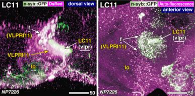 |
| Distribution of the presynaptic sites. Staining with the presynaptic site-targeted n-syb::GFP (green to white) and cytoplasmic DsRed (magenta). White characters with under bar indicate the areas of arborizations with presynaptic sites. Arborizations of the vlpr intrinsic neurons VLPRI11 are also shown (“f”). Modified from Otsuna and Ito (2006). |
| |
File 1: |  |
| QTVR movie of LC11. from Otsuna and Ito (2006). |