| Accession number: | 10012 | |
| VFB id: | FBbt_00100485   | |
| Neuron name: | LC13 |
| Synonyms: | |
| Position of cell bodies: | LCBR-LVA |
| Number of cells: | 16-17 |
| Neuron class: | visual projection neuron, lobula columnar neuron |
| Innervating regions: | ventral part of LO3, ventral part of LO4s, ventral part of LO5, PLP, ventral part of LO4d |
| Presynaptic sites: | PLP |
| Postsynaptic sites: | |
| Direction of information: | centripetal |
| Laterality: | ipsilateral |
| Publications: | -
Otsuna and Ito (2006) J. Comp. Neurol. 497:928-958 |
| |
| Strains / Antibodies: | |
| |
| Morphological description: | LC13 (Fig. 1) is the only lobula-specific columnar (LC) pathway that terminates in the plpr. We found 16–17 cell bodies that lie in the anterior ventral area of the lateral cell body region. The cell body fibers run dorsally and then bifurcate to contribute to lobula and plpr at the neck of the lobula (Fig.1, the left and the middle panels). The LC13 neurons have synaptic varicosities in the LO3, 4, and 5 layers.
Unlike other LC neurons, LC13 neurons arborize only in the ventralmost area of the lobula (Fig. 1, the left panel), where they form columnar projection running parallel along the visual cartridges. The projections from these ventral cartridges form a bundle that runs dorsally towards the lobula neck, where they form a steep turn (Fig. 1). The bundle turns medially and posteriorly at the lobula neck (Fig.1, the left and the middle panels), enters the lateral area of the plpr, and forms a cone-like terminal arborization. Presynaptic sites are observed only in the terminal end in the plpr (Fig. 1). LC13 is therefore centripetal.
(Otsuna and Ito, 2007) |
| |
| Functional description: | |
| |
| |
| Figure 1: | 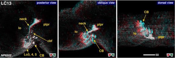 |
| 3D stereograms from three viewing angles. (Neurons other than LC13 are erased from the data. )
CB, position of the cell bodies; cbf, cell body fibers; neck, medial edge of the lobula where all the lobula-specific VPNs converge; plpr, posterolateral protocerebrum; Lo 3, 4, 5 arborized layers in the lobula. Modified from Otsuna and Ito (2006). |
| |
Figure 2: | 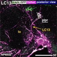 |
| Distribution of the presynaptic sites. Staining with the presynaptic site-targeted n-syb::GFP (green to white) and cytoplasmic DsRed (magenta). White characters with under bar indicate the areas of arborizations with presynaptic sites. Modified from Otsuna and Ito (2006). |
| |
File 1: |  |
| QTVR movie of LC13, from Otsuna and Ito (2006). |