| Accession number: | 10014 | |
| VFB id: | FBbt_00003860   | |
| Neuron name: | LT1 |
| Synonyms: | Lt1, LTL1 |
| Position of cell bodies: | LCBR-LDA |
| Number of cells: | 4 |
| Neuron class: | visual projection neuron, lobula tangential neuron |
| Innervating regions: | LO3, AVLP |
| Presynaptic sites: | AVLP |
| Postsynaptic sites: | LO3 |
| Direction of information: | centripetal |
| Laterality: | ipsilateral |
| Publications: | -
Fischbach and Dittrich (1989) Cell Tissue Res. 258:441-475
-
Otsuna and Ito (2006) J. Comp. Neurol. 497:928-958 |
| |
| Strains / Antibodies: | NP1195-Gal4 |
| |
| Morphological description: | LT1 consists of four neurons that have their cell bodies in the anterior dorsal area of the lateral cell body region (Figs. 5, 6). This pathway has been identified as Lt1 (lobula tangential 1) (Fischbach and Dittrich, 1989). The cell body fibers run toward the lobula neck and bifurcate to project to the lobula and the vlpr (Fig. 5). In the lobula, the fibers initially run along its medial and posterior surface (Figs. 5, the right panel; 6, the left panel). Upon reaching the posteriormost edge of the lobula, the fibers enter the lobula neuropil to form thin, tangential arborization in the LO3 layer (Fig. 5, the middle panel). A single neuron covers the whole visual field (Fig. 6, the right panel). In the vlpr, the fiber bundle first projects into the vlpr, makes a steep turn laterally (arrows in Fig. 5), and arborizes in the lateralmost vlpr to form many varicosities. The n-syb::GFP signal was observed only in the vlpr (Fig. 7), suggesting that LT1 neurons are centripetal. |
| |
| Functional description: | |
| |
| |
| Figure 1: | 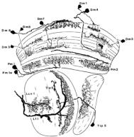 |
| Composite of camera lucida drawing, from Fischbach and Dittrich (1989), Figure 5. |
| |
Figure 2: | 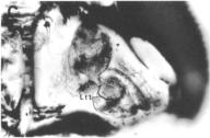 |
| Horizontal section, Golgi-Colonnier procedure, from Fischbach and Dittrich (1989), Figure 2C. |
| |
Figure 3: | 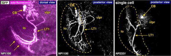 |
| Morphology of the LT1 neurons of the strains that label four (left, middle) and one (right) neurons. A pair of arrows in the left panel indicates the direction of the visual columns. Dashed lines indicate the contour of the lobula. Modified from Otsuna and Ito (2006). |
| |
Figure 4: | 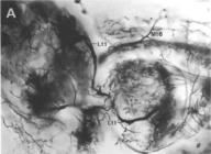 |
| From Fischbach and Dittrich (1989), Figure 28A. |
| |
Figure 5: |  |
| 3D stereograms from three viewing angles. Arrowheads indicate the edges of the terminal arborization. (Neurons other than LT1 are erased from the data. ) Arrows indicate the point where the fiber makes a steep turn. CB, position of the cell bodies; vlpr, ventrolateral protocerebrum; LO3: arborized layers in the lobula.
Modified from Otsuna and Ito (2006).
|
| |
Figure 6: | 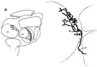 |
| From Fischbach and Dittrich (1989), Figure 22A. |
| |
Figure 7: | 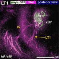 |
| Distribution of the presynaptic sites. Staining with the presynaptic site-targeted n-syb::GFP (green to white) and cytoplasmic DsRed (magenta). White characters with under bar indicate the areas of arborizations with presynaptic sites. Modified from Otsuna and Ito (2006). |
| |
File 1: |  |
| QTVR movie of LT1, from Otsuna and Ito (2006). |