| Accession number: | 10015 | |
| VFB id: | FBbt_00003869   | |
| Neuron name: | LT10 |
| Synonyms: | Lt10, LTL3 |
| Position of cell bodies: | LCBR-LDA |
| Number of cells: | 1 |
| Neuron class: | visual projection neuron, lobula tangential neuron |
| Innervating regions: | LO4_dorsal, AVLP |
| Presynaptic sites: | AVLP |
| Postsynaptic sites: | |
| Direction of information: | centripetal |
| Laterality: | ipsilateral |
| Publications: | -
Otsuna and Ito (2006) J. Comp. Neurol. 497:928-958
-
Fischbach and Dittrich (1989) Cell Tissue Res. 258:441-475
-
Strausfeld and Campos-Ortega (1977) Science 195(4281):894-897 |
| |
| Strains / Antibodies: | NP1035-Gal4, NP1047-Gal4, NP6099-Gal4 |
| |
| Morphological description: | The LT10 pathway consists of a single neuron (Fig. 1), which is likely identical with Lt10 (Fischbach and Dittrich, 1989). Its cell body is located in the anterior dorsal area of the lateral cell body region. The cell body fiber runs toward the lobula neck, from where the fiber runs dorsally to project to the dorsal part of the lobula. The fiber forms branches at the dorsalmost edge of the lobula and sends tangential arborizations dorsoventrally along the LO4 layer (Fig. 1, the middle panel). The arborization covers only the dorsal half of the visual field (Fig. 1, the left panel). Horizontally it covers the whole visual field (Figs. 1, the right panel; 2).
The fiber toward the vlpr runs along the lateral edge of the vlpr (Figs. 1, the right panel; 2) and terminates in the dorsolateral vlpr, roughly at the same area as the target of the LT1 and LT11. Presynaptic sites were observed only in the vlpr (Fig. 3), suggesting that it is a centripetal neuron.
(Otsuna and Ito, 2007) |
| |
| Functional description: | |
| |
| |
| Figure 1: | 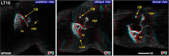 |
| 3D stereograms from three viewing angles. (Neurons other than LT10 are erased from the data.)
CB, position of the cell bodies; vlpr, ventrolateral protocerebrum; LO4, arborized layers in the lobula. Modified from Otsuna and Ito (2006). |
| |
Figure 2: | 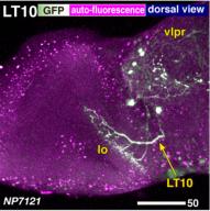 |
| Detailed arborization pattern of LT10. Modified from Otsuna and Ito (2006). |
| |
Figure 3: | 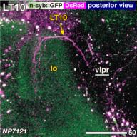 |
| Distribution of the presynaptic sites. Staining with the presynaptic site-targeted n-syb::GFP (green to white) and cytoplasmic DsRed (magenta). White characters with under bar indicate the areas of arborizations with presynaptic sites. Modified from Otsuna and Ito (2006). |
| |
Figure 4: | 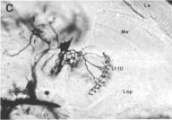 |
| From Fischbach and Dittrich (1989), Figure 28C. |
| |
File 1: | 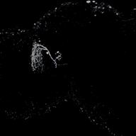 |
| QTVR movie of LT10, from Otsuna and Ito (2006). |