| Accession number: | 10016 | |
| VFB id: | FBbt_00100487   | |
| Neuron name: | LT11 |
| Synonyms: | |
| Position of cell bodies: | LCBR-LDA |
| Number of cells: | 1 |
| Neuron class: | visual projection neuron, lobula tangential neuron |
| Innervating regions: | LO3, LO4, LO5, AVLP |
| Presynaptic sites: | LO3, LO4, AVLP |
| Postsynaptic sites: | |
| Direction of information: | bidirectional |
| Laterality: | ipsilateral |
| Publications: | -
Otsuna and Ito (2006) J. Comp. Neurol. 497:928-958 |
| |
| Strains / Antibodies: | NP6099-Gal4, NP1035-Gal4 |
| |
| Morphological description: | The LT11 pathway consists of a single neuron. The cell body is located in the anterior dorsal area of the lateral cell body region (Fig. 1). The neuron has tree-like branches in the lobula and form varicosities in two separate layers. The outer (lateral) layer corresponds to the LO3 and 4, and the inner (medial) layer corresponds to LO5 (Fig. 1). In the vlpr the neuron arborizes in roughly the same anterior dorsal area as that of the LT1.
The presynaptic sites are found in the LO3 and 4 layers of the lobula as well as in the vlpr (Fig. 3). The varicosities in the LO5 layer are devoid of n-syb::GFP and are hence likely to be postsynaptic. The LT11 neuron therefore seems to collect information in the LO5 layer and transmit the signal centrifugally to the LO3, 4 layers and centripetally to the vlpr.
(Otsuna and Ito, 2007) |
| |
| Functional description: | |
| |
| |
| Figure 1: | 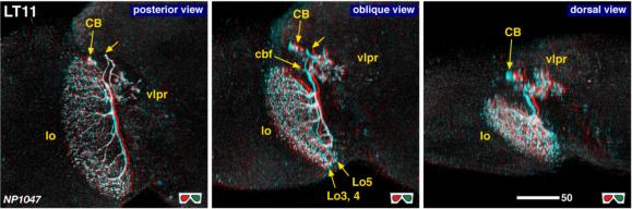 |
| 3D stereograms from three viewing angles. (Neurons other than LT11 are erased from the data.) CB, position of the cell bodies; cbf, cell body fiber; vlpr, ventrolateral protocerebrum; LO3, 4, 5, arborized layers in the lobula. Modified from Otsuna and Ito (2006). |
| |
Figure 2: | 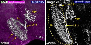 |
| Morphology of the LT11 neuron. A pair of arrows in the left panel indicates the direction of the visual columns. Dashed lines indicate the contour of the lobula. Modified from Otsuna and Ito (2006). |
| |
Figure 3: | 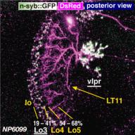 |
| Distribution of the presynaptic sites. Staining with the presynaptic site-targeted n-syb::GFP (green to white) and cytoplasmic DsRed (magenta). White characters with under bar indicate the areas of arborizations with presynaptic sites. Modified from Otsuna and Ito (2006). |
| |
File 1: |  |
| QTVR movie of LT11, from Otsuna and Ito (2006). |