| Accession number: | 10018 | |
| VFB id: | FBbt_00100489   | |
| Neuron name: | LT31 |
| Synonyms: | |
| Position of cell bodies: | brain(near plpr) |
| Number of cells: | 1 |
| Neuron class: | visual projection neuron, lobula tangential neuron |
| Innervating regions: | LO4, PLP |
| Presynaptic sites: | LO4 |
| Postsynaptic sites: | |
| Direction of information: | centrifugal |
| Laterality: | ipsilateral |
| Publications: | -
Otsuna and Ito (2006) J. Comp. Neurol. 497:928-958 |
| |
| Strains / Antibodies: | |
| |
| Morphological description: | The LT31 consists of a single neuron. The cell body lies in the central brain cortex posterior to the plpr (Fig. 1). The cell body fiber runs anteriorly straight through the plpr neuropil, and bifurcates in the vlpr (arrowhead in Fig. 1). The lateral branch makes a round turn and runs ventral-posteriorly toward the neck of the lobula. From there the neuron forms extensive tree-like branches
with varicosities confined in the LO4 layer. The medial branch makes a right-angle turn (arrow in Fig. 1) to run ventral-posteriorly towards the posterior plpr. It forms terminal arborizations in the area that is close to the position of the cell body (Fig. 1, the right panel). Although the distance between the two areas of arborization (plpr and lo) is fairly close, the neuron makes a long U-shaped detour to connect them.
The n-syb::GFP signal of LT31 was observed only in the lobula (Fig. 2). Arborization in the plpr is devoid of presynapses. There were no varicosities along the trajectory through the vlpr. The LT31 neuron is therefore centrifugal, sending information from the plpr to the LO4 layer of the lobula.
(Otsuna and Ito, 2007) |
| |
| Functional description: | |
| |
| |
| Figure 1: | 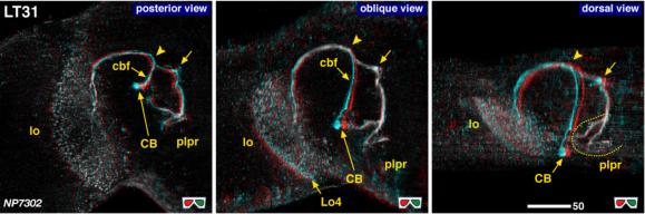 |
| 3D stereograms from three viewing angles. Arrowheads indicate the bifurcation point of the cell body fiber. (Neurons other than LT31 are erased from the data. ) Arrows indicate the point where the fiber makes a right-angled turn. CB, position of the cell bodies; plpr, posterolateral protocerebrum; LO4: arborized layers in the lobula.
Modified from Otsuna and Ito (2006). |
| |
Figure 2: | 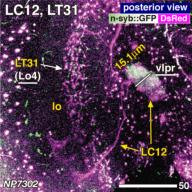 |
| Distribution of the presynaptic sites. Staining with the presynaptic site-targeted n-syb::GFP (green to white) and cytoplasmic DsRed (magenta). White characters with under bar indicate the areas of arborizations with presynaptic sites. Modified from Otsuna and Ito (2006). |
| |
File 1: |  |
| QTVR movie of LT31, from Otsuna and Ito (2006). |