| Accession number: | 10019 | |
| VFB id: | FBbt_00100490   | |
| Neuron name: | LT32 |
| Synonyms: | |
| Position of cell bodies: | brain(oesophagus) |
| Number of cells: | 1 |
| Neuron class: | visual projection neuron, lobula tangential neuron |
| Innervating regions: | LO5, LO6, contralateral LO, PLP, contralateral PLP |
| Presynaptic sites: | LO5, LO6, contralateral PLP |
| Postsynaptic sites: | |
| Direction of information: | centrifugal |
| Laterality: | inter-OL |
| Publications: | -
Otsuna and Ito (2006) J. Comp. Neurol. 497:928-958 |
| |
| Strains / Antibodies: | |
| |
| Morphological description: | The LT32 pathway also consists of a single neuron per hemisphere (Fig. 1). A pair of cell bodies lies on each side of the esophageal foramen. The cell body fiber runs posteriorly along the oesophagus, and turns upwards near the posterior end of the brain (Fig. 2, the right panel). There the fiber forms a Y-shaped branch (arrowheads in Figs. 1, the right panel; 3). One branch runs laterally to project to the lobula, and the other crosses the midline to innervate the contralateral lobula. Along the trajectory towards the lobula, the fiber forms another collateral branch (white arrows in Fig. 1), which innervates the plpr and forms fine arborizations. The main branch reaches the lobula and forms extensive tree-like arborizations with varicosities in the LO5 and 6 layers (Fig. 1, the middle panel).
We also found an interesting example of misrouting projections. In one sample the cell body fiber of one side of the LT32 turns upward prematurely before the fiber reaches the posterior end of the brain (Fig. 2, the left and the middle panel). The misrouted projection nevertheless turns again posteriorly to reach the posterior surface of the brain. The fiber forms the Y-shaped branch and innervates the plpr and lobula-like normal neurons. This suggests that the fibers of LT32 have the ability to find the correct projection target even when the fiber lost its way in the complex neuropil structure.
Because LT32 neurons on both hemispheres project along almost the same trajectory, it is difficult to identify the presynaptic sites of the neuron of a particular hemisphere. Fortunately, we found one sample in which DsRed failed to express in one of the LT32 neurons (Fig. 3). In the contralateral side of the projection (Fig. 3, the top panel), extensive distribution of presynapses was observed in the plpr as well as in the lobula. In the ipsilateral side (Fig. 3, the bottom panel), the plpr is essentially devoid of presynapses, although there are arborizations. Presynapses are observed only in the lobula. Thus, it is likely that LT32 collect information in the ipsilateral plpr and send the signal to the lobulae on both hemispheres as well as to the contralateral plpr.
(Otsuna and Ito, 2007) |
| |
| Functional description: | |
| |
| |
| Figure 1: | 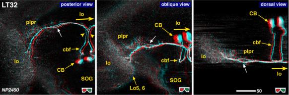 |
| 3D stereograms from three viewing angles. Arrowheads indicate bifurcation point of the cell body fiber. (Neurons other than LT32 are erased from the data. ) Arrows indicate the point where the fiber makes a right-angle turn. CB, position of the cell bodies; plpr, posterolateral protocerebrum; SOG, suboesophageal ganglion; LO5, 6,: arborized layers in the lobula.
Modified from Otsuna and Ito (2006). |
| |
Figure 2: | 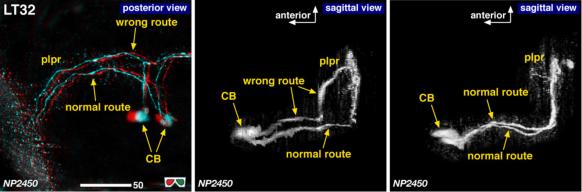 |
| Detailed arborization pattern of LT32. Left and middle: A sample in which LT32 of one hemisphere took a wrong route to innervate. Right: Normal route of LT32.
Modified from Otsuna and Ito (2006). |
| |
Figure 3: | 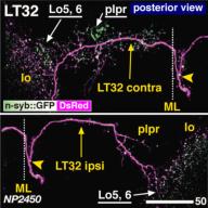 |
| Distribution of the presynaptic sites. Staining with the presynaptic site-targeted n-syb::GFP (green to white) and cytoplasmic DsRed (magenta). White characters with under bar indicate the areas of arborizations with presynaptic sites. Modified from Otsuna and Ito (2006). |
| |
File 1: |  |
| QTVR movie of LT32, from Otsuna and Ito (2006). |