| Accession number: | 10365 | |
| VFB id: | FBbt_00003994   | |
| Neuron name: | GSI |
| Synonyms: | giant symmetric relay interneurons |
| Position of cell bodies: | ventral midline of SOG |
| Number of cells: | |
| Neuron class: | olfactory secondary neuron |
| Innervating regions: | AL |
| Presynaptic sites: | |
| Postsynaptic sites: | |
| Direction of information: | |
| Laterality: | |
| Publications: | -
Stocker et al. (1990) Cell Tissue Res. 262:9-34 |
| |
| Strains / Antibodies: | |
| |
| Morphological description: | Giant symmetric relay interneurons (GSI), observed in two preparations, are very conspicuous because of their large size and their extensive symmetric arborization in both antennal lobes. The cell body of these neurons is located in the midline of the SOG (suboesophageal ganglion), i.e., in its anteroventral cortex. A stout process extends dorsally from the cell body through the SOG neuropil and splits below the oesophagus into a bilateral pair of similarly large processes. Close to the branching point, the two processes give rise to a mirror-symmetric pattern of arborizations on both sides of the oesophagus. Some branches even extend into posterior brain regions behind the great commissure. The two main processes turn abruptly into the anterior direction to reach the antennal lobes through the broad root (Power (1946) J. Morphol. 72:517-559), just ventral to the iACT (inner antenno-cerebral tract). They proceed into the center of the lobe and send off a large number of secondary and tertiary processes that extend into the periphery of the lobe. Virtually all of the glomeruli appear to be reached by these branches; in addition, terminal branches seem to extend some distance into the antennal nerve. With respect to the branching pattern, two subtypes of GSIs have been found. In the first one, branches are uniformly thin, whereas, in the second one, thick and thin branches are formed: the thick branches occupy approximately the antero-lateral half of the lobe, whereas the fine branches cover the rest. Moreover, in the second type, the additional branching region in the posterior brain apparently does not reach the region of the great commissure. |
| |
| Functional description: | |
| |
| |
| Figure 1: | 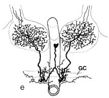 |
| From Stocker et al. (1990), Figure 14e. |
| |
Figure 2: | 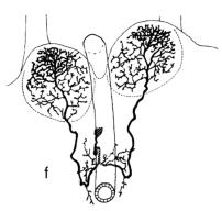 |
| From Stocker et al. (1990), Figure 14f. |
| |
Figure 3: | 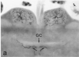 |
| Dorsal aspect of GSI. From Stocker et al. (1990), Figure 16a. |
| |
Figure 4: | 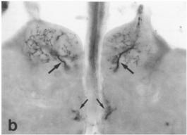 |
| Middle aspect of GSI. From Stocker et al. (1990), Figure 16b. |
| |
Figure 5: |  |
| Ventral aspect of GSI. From Stocker et al. (1990), Figure 16c. |