| Accession number: | 10367 | |
| VFB id: | FBbt_00003996   | |
| Neuron name: | TI |
| Synonyms: | thoracic relay interneuron |
| Position of cell bodies: | antero-dorsal cortex of brain |
| Number of cells: | |
| Neuron class: | olfactory secondary neuron |
| Innervating regions: | ventral half of AL, thoracic ganglion, PLP? [posterior lateral brain in Stocker et al. (1990)] |
| Presynaptic sites: | |
| Postsynaptic sites: | |
| Direction of information: | |
| Laterality: | |
| Publications: | -
Stocker et al. (1990) Cell Tissue Res. 262:9-34 |
| |
| Strains / Antibodies: | |
| |
| Morphological description: | Thoracic relay interneuron (TI) has been found that arborizes profusely in a number of antennal glomeruli, i.e., in VA3, DL1/DA3, and in the VP1-3 region. These arborizations, originate from a stout process that leaves the antennal lobe through the AST (antenno-suboesophageal tract). Shortly behind the lobe, the process sends off a very long fiber towards the cell body, which is located in the antenno-dorsal cortex of the lobe, near the midline. The main process divides once more. One branch extends into the posterior brain and establishes terminal arborizations just behind the great commissure; the other branches project thorough the SOG and neck connective into the thoracic ganglion. Its thoracic targets are not known. |
| |
| Functional description: | |
| |
| |
| Figure 1: | 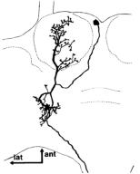 |
| From Stocker et al. (1990), Figure 17. |