| Accession number: | 10535 | |
| VFB id: | FBbt_00050149   | |
| Neuron name: | clonal unit CREa1 |
| Synonyms: | |
| Position of cell bodies: | raCRE |
| Number of cells: | 107 |
| Neuron class: | clonal unit |
| Innervating regions: | |
| Presynaptic sites: | |
| Postsynaptic sites: | |
| Direction of information: | |
| Laterality: | |
| Publications: | |
| |
| Strains / Antibodies: | |
| |
| Morphological description: | raCRE-<raCRE>{
-<CRE>{
-|CRE-MLF|-o[ML]-|MLC|-o[ML']/
-|CRE-SMPF|-<SMP>{
-[CRE+SIP+SMP+SLP]/
-[SMP+SLP]}}/
-|AL-LALF|-|LALC|-<LAL'>{
-|LAL-FLAF'|-[FLA'+SAD'+SEG]/
-|LAL-ALF'|-|AL-SLPF'|-o[SLP']}}
|
| |
| Functional description: | |
| |
| |
| Figure 1: | 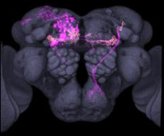 |
| Reconstruction of the clonal unit (anterior view).
Magenta : cell bodies and neuronal fibers
White : distributions of presynaptic sites
Gray : the entire neuropil of the template brain |
| |
Figure 2: | 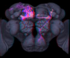 |
| Reconstruction of the clonal unit (anterior view). 3D stereogram version. Depth information can be obtained when the images are viewed through red-cyan stereo glasses (red: left eye, cyan: right eye).
Magenta : cell bodies and neuronal fibers
White : distributions of presynaptic sites
Gray : the entire neuropil of the template brain |
| |
Figure 3: |  |
| Download raw confocal stack data of the clonal unit
(TIFF serial images, ZIP compressed, not morphed into the template brain.)
Red channel : cell bodies and neuronal fibers (cytoplasmic UAS-DsRed / UAS-GFP)
Green channel : presynaptic sites (synaptic vesicle-targeted UAS-n-syb-GFP / UAS-syt-GFP)
Blue channel : nc82 labeling of neuropils |