| Accession number: | 10001 | |
| VFB id: | FBbt_00003874   | |
| Neuron name: | LC4 |
| Synonyms: | Lcn4 |
| Position of cell bodies: | LCBR-LV |
| Number of cells: | 25-29 |
| Neuron class: | visual projection neuron, lobula columnar neuron |
| Innervating regions: | LO2, LO4, PVLP (GL10) [vlpr in Otsuna and Ito (2006), Fischbach and Dittrich (1989)] |
| Presynaptic sites: | PVLP (GL10) [vlpr in Otsuna and Ito (2006), Fischbach and Dittrich (1989)] |
| Postsynaptic sites: | LO2, LO4 |
| Direction of information: | centripetal |
| Laterality: | ipsilateral |
| Publications: | -
Otsuna and Ito (2006) J. Comp. Neurol. 497:928-958
-
Fischbach and Dittrich (1989) Cell Tissue Res. 258:441-475 |
| |
| Strains / Antibodies: | NP7476-Gal4, NP7281-Gal4, NP5039-Gal4, NP2308-Gal4, NP2469-Gal4 |
| |
| Morphological description: | LC4 neurons have previously been identified as lobula columnar neurons 4 (Lcn4) (Fischbach and Dittrich, 1989). 25-29 cell bodies (CBs) were observed in the mid-ventral area of the lateral cell body region. The cell body fibers (cbf) form a bundle that runs towards the “neck” of the lobula, which is the medial edge of this neuropile where all the lobula-specific VPNs converge. At the neck, the fibers bifurcate to contribute to the lobula and to the ventrolateral protocerebrum (vlpr).
In the lobula they form clear columnar projections that cover the whole visual field. Whereas the medial half of the lobula contains only thick branches, the lateral half is rich in numerous fine arborizations, with bi-stratified varicosities in layers LO2 and LO4.
In the central brain, the fibers form a thick prominent bundle called the “lobula/lobula plate bundle”, which terminates in the posteriormost medial area of the vlpr. These terminals form a blunt, stick-like ending.
Single neurons of this pathway send fibers along the visual cartridges. Right panel of Figure 3 shows two such VPNs that arborize in different visual cartridges. Detailed examination of confocal sections revealed that the two axons intertwine as they project to the vlpr but eventually arborize in a separate area of the vlpr (schematic drawing in Figure 3). This would suggest that some sort of retinotopic spatial map is maintained between the lobula and the vlpr.
The n-syb::GFP labeling was observed only in the medial half of the stick-like ending in the vlpr (Figure 4). Single-neuron staining clearly shows varicosities in this area (Figure 3). LC4 is therefore centripetal. Branches in the lobula, which are not labeled with n-syb::GFP, also show varicosity-like structures (arrowheads in Figure 3 right panel), although the size of these blebs is relatively small.
Both LC4 and LC11 have extensive columnar arborizations in the lobula. Their varicosities overlap in LO2 and LO4 layers. Yet, their projection targets in the vlpr are strikingly different. Terminals of LC11 are located relatively dorsally, whereas LC4 terminate more ventrally. LC4 enters the vlpr from its lateral side and terminate in the posteriormost area. LC11, on the other hand, enters the vlpr from the posterior side and arborizes in the lateral and anterior areas. These suggest that apparently similar set of information from the lobula is conveyed to different subregions of the vlpr.
(Otsuna and Ito, 2007)
|
| |
| Functional description: | |
| |
| |
| Figure 1: | 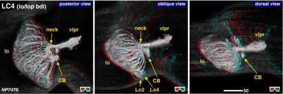 |
| 3D stereograms from three viewing angles. (Neurons other than LC4 are erased from the data.)
CB, position of the cell bodies; neck, medial edge of the lobula where all the lobula-specific VPNs converge; vlpr, ventrolateral protocerebrum; LO2, 4, arborized layers in the lobula. Modified from Otsuna and Ito (2006).
|
| |
Figure 2: | 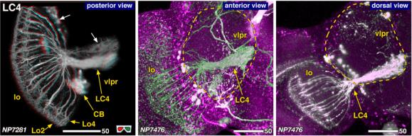 |
| Detailed arborization pattern of LC4. White arrows in the left panel indicate the cell bodies and the projection targets of the columnar VPNs arborizing lobula and lobula plate (CC VPNs). Dashed lines in the mid and right panels indicate the contour of the vlpr. Modified from Otsuna and Ito (2006). |
| |
Figure 3: | 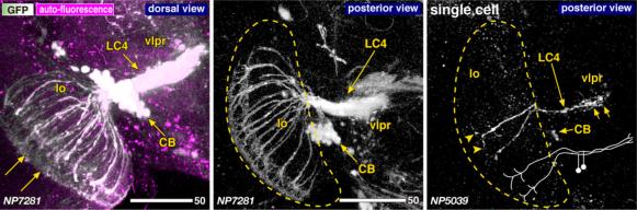 |
| Morphology of the LC4 neurons of the strains that label most (left, middle) and only a few (right) neurons. A pair of arrows in the left panel indicates the direction of the visual columns. Dashed lines indicate the contour of the lobula. Drawing in the right panel shows the schematics of the two neurons labeled in this sample. Modified from Otsuna and Ito (2006). |
| |
Figure 4: | 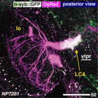 |
| Distribution of the presynaptic sites. Staining with the presynaptic site-targeted n-syb::GFP (green to white) and cytoplasmic DsRed (magenta). White characters with under bar indicate the areas of arborizations with presynaptic sites. Modified from Otsuna and Ito (2006). |
| |
Figure 5: | 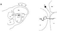 |
| Camera lucida drawing of the Golgi-impregnated preparation. From Fischbach and Dittrich (1989), Figure 23A. |
| |
File 1: | 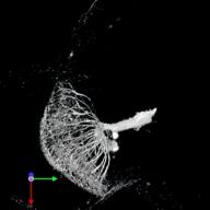 |
| QTVR movie of LC4. from Otsuna and Ito (2006). |