| Accession number: | 10003 | |
| VFB id: | FBbt_00003876   | |
| Neuron name: | LC6 |
| Synonyms: | AOT, Lcn6, S4 |
| Position of cell bodies: | LCBR-LD |
| Number of cells: | ~150? |
| Neuron class: | visual projection neuron, lobula columnar neuron |
| Innervating regions: | PVLP (GL2) [vlpr in Otsuna and Ito (2006), Fischbach and Dittrich (1989)] |
| Presynaptic sites: | PVLP (GL2) [vlpr in Otsuna and Ito (2006), Fischbach and Dittrich (1989)] |
| Postsynaptic sites: | |
| Direction of information: | centripetal |
| Laterality: | ipsilateral |
| Publications: | -
Otsuna and Ito (2006) J. Comp. Neurol. 497:928-958
-
Fischbach and Dittrich (1989) Cell Tissue Res. 258:441-475
-
Fischbach (1983) Dev. Biol. 95(1):1-18 |
| |
| Strains / Antibodies: | NP6250-Gal4 |
| |
| Morphological description: | LC6 forms the anterior optic tract (AOT; Power, 1943) together with LC9 and LC10. They are described as Lcn6, 9 and 10 in Fischbach and Dittrich (1989).
The cell bodies of LC6, 9 and 10 form a single large cluster in the mid dorsal area of the lateral cell body region. Like all other LCs, the cell body fibers run towards the lobula neck and bifurcate there.
The AOT is a prominent fiber bundle that runs from the lobula to the optic tubercle (optu). Whereas LC10 runs straight and projects to the optu, LC6 and 9 make a round turn in the midway of the AOT to terminate in the vlpr. The bundles of LC6/9 and LC10 are separated clearly in the AOT. The semicircular trajectory in the vlpr is characteristic to LC6/9. They are also the only VPNs that enter the vlpr from its anterior side.
Neurons in the AOT have been classified into four types: S1-S4 (Fischbach and Lyly-Hünerberg, 1983). S1 neurons connect different regions of the central brain, and the origin and the projection target of S2 neurons are not described in enough detail. The relatively straight trajectory of the S3 neurons and the round trajectory of the S4 axons indicate that they correspond to the LC10 and LC6/9 pathways, respectively.
The bundles of LC6 and LC9 run together along the AOT. Their trajectories separate only after they enter the vlpr. LC6 forms a steeper turn than LC9 and terminate in the lateralmost area of the dorsal posterior vlpr. The distal end of the bundle spreads horizontally to form a triangular shape that spans from the central vlpr to its lateral edge (arrowheads in Figure 1 and Figure 3 left panel).
In the lobula, LC6 arborizes in the LO3, 4, 5 and 6 layers. The n-syb::GFP signal in the lobula was observed in a thin layer in LO6 (Figure 4). In the vlpr, the output sites are confined in the triangular area. LC6 is therefore a centripetal pathway sending information from LO3-5 layers to the vlpr, with collateral output to the LO6 layer.
We found no GAL4 strains that specifically label the LC6 neurons. LC6, 9 and 10 neurons are labeled collectively with the NP0681 and NP7067 strains. NP2620, on the other hand, label LC4, 6 and 14. The total cell count in the cluster of the LC6, 9 and 10 neurons was 321-358 (average 333). The numbers of the LC9 and LC10 neurons were likely to be slightly less than 83-102 (average 94) and 84-90 (average 87), respectively. The difference, on average slightly more than 152 cells, could be attributed to the LC6 neurons. The number of the LC6 neurons can also be estimated in another way. The strain NP6250 labels LC9 and 10 neurons strongly but LC6 only faintly. If we assume that most of the cells counted in this strain (151-216, average 190) belongs to LC9 and 10, the difference between this cell count and the total cell count of the LC6, 9 and 10 neurons, i.e. slightly more than 143 on average, could be attributed to the LC6 neurons. Thus, two lines of evidences suggest that the cell number of the LC6 would be around 150.
(Otsuna and Ito, 2007)
|
| |
| Functional description: | |
| |
| |
| Figure 1: | 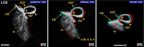 |
| 3D stereograms from three viewing angles. Arrowheads indicate the edges of the terminal arborization. (Neurons other than LC6 are erased from the data. ) Yellow arrowheads indicate the edges of the triangular projection terminals in the vlpr.
CB, position of the cell bodies; neck, medial edge of the lobula where all the lobula-specific VPNs converge; vlpr, ventrolateral protocerebrum; AOT, anterior optic tract, LO3, 4, 5, 6: arborized layers in the lobula.
Modified from Otsuna and Ito (2006).
|
| |
Figure 2: | 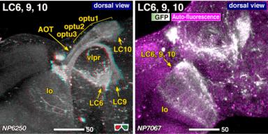 |
| Combined staining of LC6, 9 and 10, showing the spatial relationship of each pathway. optu1, 2, 3, three zones of the optic tubercle. Modified from Otsuna and Ito (2006). |
| |
Figure 3: | 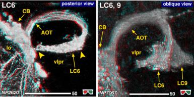 |
| Detailed arborization pattern in the vlpr. Modified from Otsuna and Ito (2006). |
| |
Figure 4: | 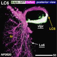 |
| Distribution of the presynaptic sites. Staining with the presynaptic site-targeted n-syb::GFP (green to white) and cytoplasmic DsRed (magenta). White characters with under bar indicate the areas of arborizations with presynaptic sites. Modified from Otsuna and Ito (2006). |
| |
Figure 5: | 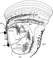 |
| Composite of camera lucida drawing. From Fischbach and Dittrich (1989), Figure 17. |
| |
Figure 6: | 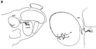 |
| Camera lucida drawing of the Golgi-impregnated preparation. From Fischbach and Dittrich (1989), Figure 17. |
| |
File 1: | 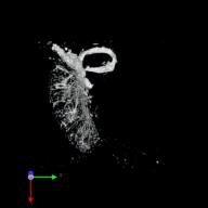 |
| QTVR movie of LC6. from Otsuna and Ito (2006). |
| |
File 2: | 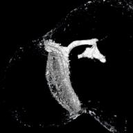 |
| QTVR movie of LC6 and 9 combined. from Otsuna and Ito (2006). |